Metrics
2024 IMPACT FACTOR
5 year Impact Factor
Eigenfactor Score
2024 CiteScore
Journal Citation Reports
(Clarivate 2025, JIF Rank)

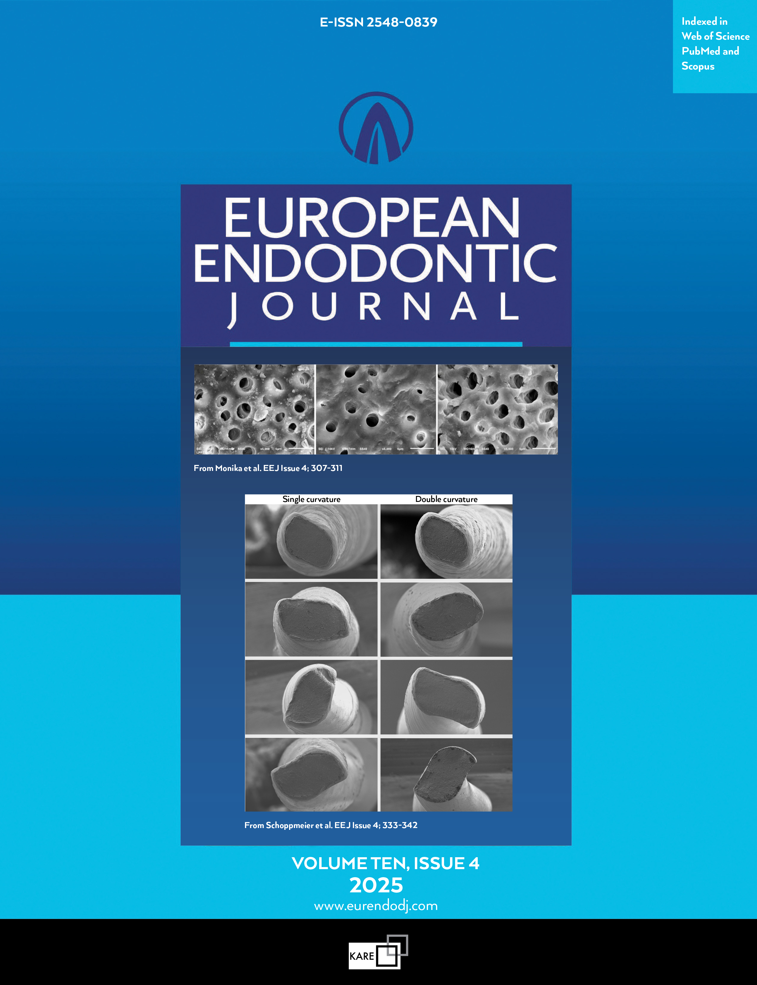
Radiomic Parameters in Periapical Lesions: A CBCT Analysis Evaluating Volumetric Size, Cortical Expansion, Erosion, and Shape
Óscar Lozano Gonzalez1, Marco Felipe Salas Orozco1, Jaime Trigueros Mancera2, Noé Gustavo Martínez Cuellar3, Nuria Patińo Marín11Department of Clinical Research, Autonomous University of San Luis Potosí, Faculty of Stomatology, San Luis Potosí, México2Department of Endodontics, Latin University, Guanajuato, Mexico
3Department of Endodontics, University of Guadalajara, Jalisco, Mexico
Objective: To investigate significant differences in selected radiomic parameters when classifying periapical lesions based on volumetric size, cortical expansion, erosion, and shape using Cone Beam Computed Tomography (CBCT).
Methods: A retrospective analytical and comparative study was conducted on 100 small field of view (FOV) 50×50 mm CBCT scans collected between the years 2018 and 2023. The study involved qualitative classification of periapical lesions, followed by segmentation and extraction of radiomic parameters. The extracted parameters included first-order features such as energy, entropy, total energy, and uniformity; texture features like grey-level co-occurrence matrix contrast (GLCMC) and neighbouring grey tone difference matrix contrast (NGTDMC); and shape features including elongation, flatness, sphericity, and mesh volume, utilising 3D Slicer and Pyradiomics. The normal distribution of the variables was determined using the Shapiro-Wilk test. Various tests were used to assess significant differences, including Student’s t-test, Mann-Whitney U test, ANOVA, and Tukey’s post hoc analysis.
Results: Significant differences were observed in the following parameters among the classification levels when classifying periapical lesions according to their volumetric size. There were significant differences in energy with a p-value of 0.001 and total energy with a p-value of 0.02. NGTDMC also showed a significant difference with a p-value of 0.001. A larger volumetric size is associated with greater energy and lower contrast. Significant differences in periapical lesions with erosion were found in shape sphericity (mean 0.34, SD 0.10, p=0.01), energy (mean 3.73×10ą⁰, SD 4.52×10ą⁰, p=0.002), and NGTDMC (mean 0.05, SD 0.02, p=0.001) compared to lesions without erosion. GLCMC was lower in erosive lesions (mean 18.94, SD 6.81, p=0.03) than in non-erosive ones (mean 22.28, SD 8.48). Regular-shaped periapical lesions demonstrated significantly greater elongation (mean 0.794, SD 0.115, p=0.006) and flatness (mean 0.614, SD 0.107, p=0.005) than irregular-shaped lesions. These findings suggest that regular-shaped periapical lesions are more elongated and flatter than irregular ones. No significant differences were found in radiomic features depending on the presence or absence of expansion in the periapical lesion.
Conclusion: There are significant differences in texture and first-order radiomic features in periapical lesions classified based on size, erosion, and shape. This research's relevance lies in its potential to improve the quantitative characterisation of periapical lesions, leading to an objective interpretation. (EEJ-2023-11-159)
Manuscript Language: English
(271 downloaded)