Metrics
2024 IMPACT FACTOR
5 year Impact Factor
Eigenfactor Score
2024 CiteScore
Journal Citation Reports
(Clarivate 2025, JIF Rank)

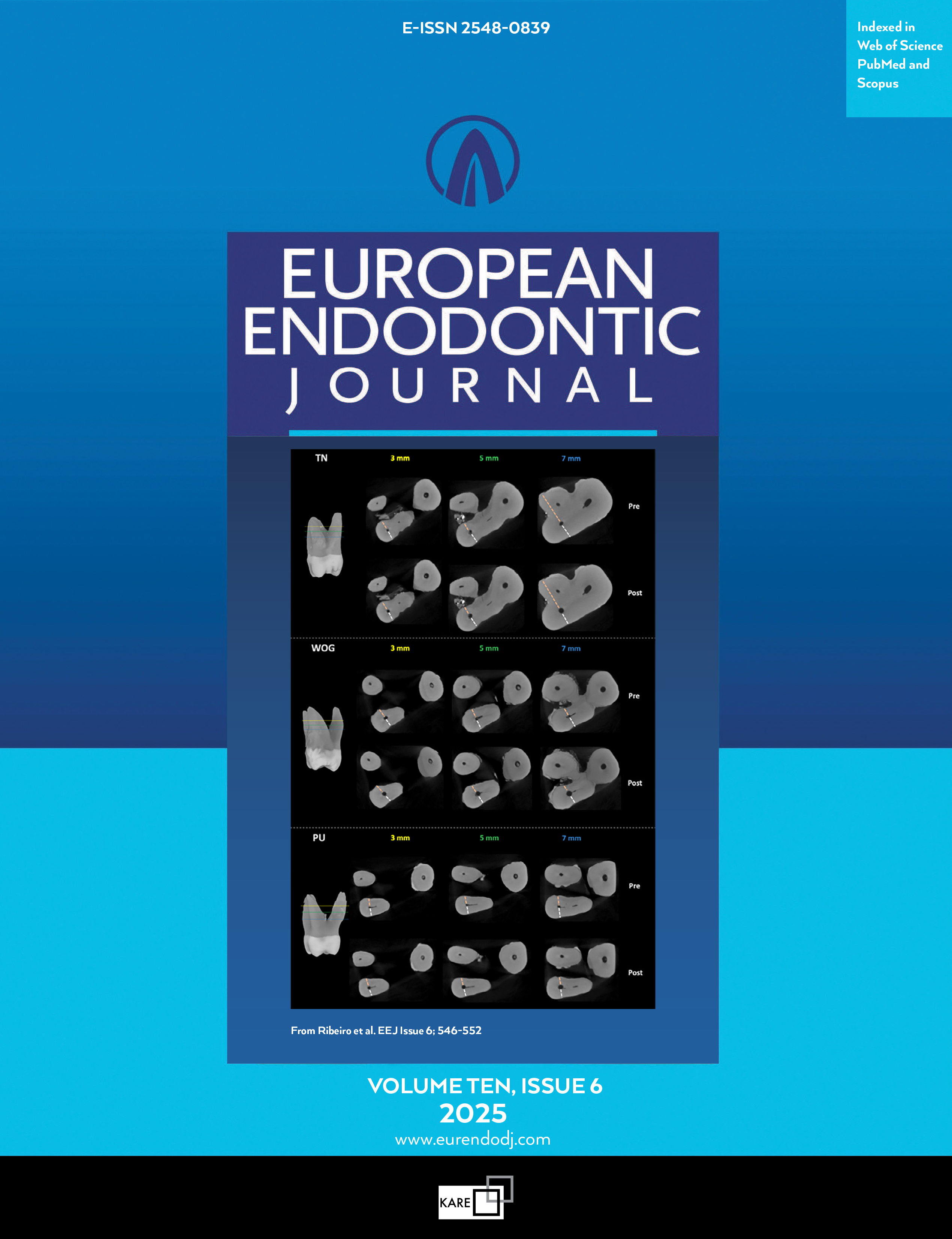
Effectiveness of a 3D Printed Training Kit for the Preparation of Access Cavities in Calcified Teeth: A Pilot Study
Vranine Kadrija1, Hauke Hildebrand1, Wadim Leontiev1, Eva Magni1, Florian Markus Thieringer2, Roland Weiger1, Thomas Connert11Department of Periodontology, Endodontology and Cariology, University Center for Dental Medicine Basel UZB, University of Basel, Basel, Switzerland2Oral and Cranio-Maxillo-Facial Surgery and 3D Print Lab, University Hospital Basel, Basel, Switzerland; Department of Biomedical Engineering, Medical Additive Manufacturing Research Group (Swiss MAM), University of Basel, Allschwil, Switzerland
Objective: This study aimed to evaluate the effectiveness of a 3D printed training kit for the preparation of endodontic access cavities in calcified teeth.
Methods: The root canal system of a micro-CT scanned premolar was digitally processed to create an endodontic training kit containing 10 teeth with ten different progressive degrees of pulp canal calcification. A tooth variant with a medium calcification degree (5/10) was printed in three copies using opaque resin. Additionally, a set of 10 transparent training teeth with red-colored pulp was produced using PolyJet 3D printing technology, which was used to train the access cavity preparation in a controlled manner due to the transparency of the teeth. Undergraduate students (n=27) and dentists (n=10) each prepared a total of 13 (one pre-training, two post-training) access cavities. Substance loss was quantified by CBCT, and user satisfaction was evaluated by questionnaire. Paired t-tests were used to compare the means for substance loss and procedure time for pre- and post-training conditions. Unpaired t-tests were used to compare differences between students and dentists. The level of significance was set at α=0.05.
Results: Mean substance loss before and after training decreased for both students (71.4 versus 54.68 mm3; p=0.069) and dentists (67.3 versus 51.1 mm3; p=0.633), but the difference was not statistically significant. The average preparation time decreased with training for students (420 versus 275 seconds; p=0.100) and dentists (336 versus 158 seconds; p=0.054), but not significantly. Root perforation rates also decreased (students: 6/27 versus 4/27; dentists: 1/10 versus 0/10). Participants rated the training model as very realistic and useful, despite the difference in material texture.
Conclusion: The proposed 3D printed training kit appears to be a suitable tool for undergraduate dental students, which could expand their opportunities to practice the preparation of endodontic access cavities in calcified teeth. (EEJ-2024-06-091)
Keywords: Endodontic education, pulp canal calcification, pulp obliteration, substance loss
Manuscript Language: English
(533 downloaded)